
brain injury
In an ‘open head injury’, brain damage due to head injury may be caused by direct intrusion into the skull by a weapon, bullet or bone fragment.

'Closed head injuries' are the result of a combination of relative movement of the brain within the skull, due to the angular rotation and/or acceleration/deceleration forces, such as may be encountered in road traffic collisions, falls or assault.
Morphological markers of brain damage include contusions and lacerations (tearing of the brain substance).
contusions
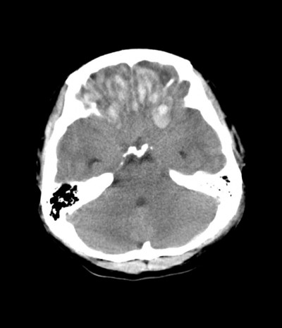
bifrontal contusions (from Radiopedia.org case 7149 Dr Frank Gaillard)
Cerebral cortical contusions are multi-focal punctate or 'streak' haemorrhages associated with foci of necrosis, which tend to be distributed along the crests of gyri. Contusions merge over time to form wedge-shaped haemorrhages with their base at the cortical surface and their apex pointing into the sub-cortical white matter.
Contusions can be classified as follows;
1. Contusions dependent upon the status of the head
- coup contusions - these occur beneath the point of impact, when the resting (but movable) head is struck by a blunt instrument. 'Pure coup' contusions are rare, and when there is a skull fracture at the site of impact, these contusions are a combination of coup and 'fracture contusions'.
- contrecoup contusions - these occur when the moving head strikes a firm surface, and the contusions are located at a point directly opposite the impacting surface. In the abscence of a skull fracture, there will be no contusion under the impacting surface. Contrecoup contusions usually develop at the frontal and temporal poles, as well as the surface of the frontal lobes, but rarely at the occipital poles. A fall onto the back of the head may therefore result in contusions of the frontal lobes etc.
- intermediate contusions - these occur in a location intermediate to coup and contrecoup contusions, and result when the moving head impacts above the level of the tentorium. They may be seen in the deep parenchyma and may mimic spontaneous intra-cerebral haemorrhages. They are usually associated with stretching injuries of the corpus callosum and are often associated with contusions in the tegmentum of the rostral pons.

Coup and contrecoup contusions - illustration of mechanism
(NB. contrecoup injuries at the back of the brain are less common than at the front of the brain, following an impact at the back of the head)
Source: Wikipedia
2. Contusions independent of the status of the head
- fracture contusions - caused by movement of bone at the site of a skull fracture
- herniation contusions - caused by the transient displacement of brain against a dural reflection, for example at the unci and cerebellar tonsils
- Gliding contusions – these are linear or oval haemorrhages within the white matter of the first frontal gyri, caused by anterior-posterior ‘gliding’ movements of the brain.
3. Blows to the resting immobile head
- Blows to the supported head may produce contusions beneath the impact site, opposite it or at an intermediate location. Shattering of the skull may accompany such an injury
'Moving head injury' contusions

Inferior frontal and temporal lobe (contre-coup) contusions (typically seen following a fall onto the back of the head)

Coronal section through contused frontal lobe

Old healed frontal and temporal contusions (with yellow edges) after fixation
Source: State University Campinas, Portugal
